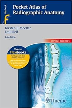
Pocket Atlas Of Radiographic Anatomy: . Zus.-Arb.: Torsten B. Möller, Emil Reif Translated By John Grossman 243 Illustrations (Thieme Flexibooks) Download Free (EPUB, PDF)

In this easily accessible pocket atlas, two expert radiologists present the normal radiographic anatomy readers need in order to interpret conventional diagnostic radiographs. Each practical, two-page unit displays a standard radiograph of a different projection on the left-hand side supplemented by a detailed, clearly labeled schematic drawing on the opposing page. The consistent, user-friendly format facilitates easy identification and rapid review of key anatomic information.Features:177 radiographic studies provide multiple views of every basic anatomic structure High-resolution radiographs appear beside explanatory drawings to aid comprehension Seven examinations new to this edition cover a trans-scapular "Y" view of the shoulder; 45° external and internal rotation views of the knee; and moreAn ideal reference for anyone involved in the interpretation of commonly performed radiographic studies, the third edition of Pocket Atlas of Radiographic Anatomy is an especially valuable tool not only for medical students and radiology residents, but also for radiological technologists.

Series: Thieme Flexibooks
Paperback: 400 pages
Publisher: TPS; 3rd edition edition (June 9, 2010)
Language: English
ISBN-10: 3137842034
ISBN-13: 978-3137842033
Product Dimensions: 5.1 x 0.9 x 7.5 inches
Shipping Weight: 1.3 pounds (View shipping rates and policies)
Average Customer Review: 4.2 out of 5 stars See all reviews (8 customer reviews)
Best Sellers Rank: #523,816 in Books (See Top 100 in Books) #97 in Books > Textbooks > Medicine & Health Sciences > Reference > Atlases #120 in Books > Textbooks > Medicine & Health Sciences > Medicine > Clinical > Radiology & Nuclear Medicine > Diagnostic Imaging #124 in Books > Medical Books > Medicine > Reference > Medical Atlases

While not useless, this book has several deficiencies that the serious clinician should consider before purchase: 1) the book is small (an advantage if one intends to keep it in a pocket or bag), which means that the images are small -- imagine trying to distinguish small details on an A-P lumbosacral spine view that measures 2" x 6". You CAN see the major features, but the smaller components are now rendered too small to see. 2) Despite the other reviews, image quality throughout this book is marginal. Yes, major structures are discernable, but subtle details (often the most important factors in noting early pathology or subtle injury) cannot be seen. The lateral cervical view in this book does not, for instance, show a prevertebral soft tissue shadow, and the spinolaminal line is barely discernable. All images are similarly affected -- the images are comparable to reading old-fashioned copy films rather than originals. In addition, some views are excessively collimated, such as the A-P cervical view which fails to show the full extent of the transverse processes of C7 --an important feature in evaluating thoracic outlet syndromes. 3) With few exceptions, the images in this text are of adults, not children (who have dramatically different normal radiographic features). 4) Although all views are named, no diagrams or instructions are given to indicate how a particular view was obtained. Some may argue that this text is not meant to be a positioning manual, but view names are not necessarily universal, making the task of requesting a particular view based on this text difficult, especially since this text originates from Germany. For example, a "Lauenstein" view of the hip joint is demonstrated. Most American x-ray techs have probably not heard of this term, making request of this view difficult unless one already recognizes that this is a lateral (frog) view of the hip joint. No text can possibly give all alternate terms for a view, which is why a small diagram illustrating patient positioning would make it much easier for a clinician to order a study, in any language, patterned after one demonstrated in this text.In my opinion, the shortcomings of this text are severe enough to exclude it from serious consideration.
I bought this because there doesn't seem to be any smaller books on the market that are just made for radiographic anatomy. Our exams include radiographic critique and many times we are asked to identify anatomy that hasn't been identified in our regular book. I thought this would be a great supplemental book. It does include a large variety of exam types, and I like that there's a drawing of the exact radiographs with anatomy pointed out rather than lines or arrows on the actual radiograph. The quality of the radiographs is pretty awful. Many of them are full of noise (fog) and/or underexposed. Makes it hard to see anatomy when it's not a high quality diagnostic image.
this is a good purchase. Recommended for those currently going through clinical rotation through a radiology program. It helped me all the way until my second year of employment. this will not fail you! small price to pay for outstanding work performance!
My go-to atlas for radiographic anatomy. Love the drawn picture for each radiograph. Good variation of views for each body part (AP, lat, oblique). I am buying more of these books in this series now to use during my radiology residency.
The lost art of the plain radiograph is kept alive in this book. Organized by standard radiographic views, this book will be useful for clinicians, as well as junior residents in radiology. The images are arranged with the radiograph on one side with the schematic and key on the other.Quality of the images are decent and some subtle soft tissue anatomy is poorly visualized but the schematic is useful. This book should be upgraded into the modern imaging techniques of digital radiography to outline the soft tissue structures.The value of contrast enhanced studies are useful: angiographic, bronchographic, lymphographic and enteric anatomy is well defined. It could use more specific terminology, especially in gastrointestinal anatomy such as defining the Z line, B line, etc.Despite some minor limitations, the book is very good and I recommend this for the framework of interpreting plain radiographs.
If you are just staring out in radiography, then this is the book you want.
love it and usefulexcellent quality, still being used like new 2 years later, i would recommened this productthanks
This book is great for the students that we teach at the hospital it has very decent pictures and is just a great all around book for xray
Pocket Atlas of Radiographic Anatomy: . Zus.-Arb.: Torsten B. Möller, Emil Reif Translated by John Grossman 243 Illustrations (Thieme Flexibooks) Atlas of Acoustic Neurinoma Microsurgery: . Zus.-Arb.: Mario Sanna Essam Saleh, Benedict Panizza, Alexandra Russo, Abdel TaibahWith the collaboration of Refik Caylan, Fernando Mancini ... Head and Neck Anatomy for Dental Medicine (Thieme Atlas of Anatomy) Atlas of Anatomy (Thieme Anatomy) Don Quixote: Translated by Edith Grossman Grant's Atlas of Anatomy (Grant, John Charles Boileau//Grant's Atlas of Anatomy) Ear Acupuncture: A Precise Pocket Atlas, Based on the Works of Nogier/Bahr (Complementary Medicine (Thieme Paperback)) Pocket Atlas of Tongue Diagnosis: With Chinese Therapy Guidelines for Acupuncture, Herbal Prescriptions, and Nutri (Complementary Medicine (Thieme Paperback)) Florida Driver's Handbook translated to Russian: Florida Driver's Manual translated to Russian (Russian Edition) Principles of Radiographic Imaging: An Art and A Science (Carlton,Principles of Radiographic Imaging) Radiographic Imaging and Exposure, 4e (Fauber, Radiographic Imaging & Exposure) Fundamentals of Special Radiographic Procedures, 5e (Snopek, Fundamentals of Special Radiographic Procedures) Stefan Grossman's Early Masters of American Blues Guitar: Mississippi John Hurt, Book & CD Atlas of Normal Radiographic Anatomy and Anatomic Variants in the Dog and Cat, 1e Anatomy: A Photographic Atlas (Color Atlas of Anatomy a Photographic Study of the Human Body) McMinn and Abrahams' Clinical Atlas of Human Anatomy: with STUDENT CONSULT Online Access, 7e (Mcminn's Color Atlas of Human Anatomy) Color Atlas of Anatomy: A Photographic Study of the Human Body (Color Atlas of Anatomy (Rohen)) Anatomy: A Regional Atlas of the Human Body (ANATOMY, REGIONAL ATLAS OF THE HUMAN BODY (CLEMENTE)) Young House Love: 243 Ways to Paint, Craft, Update & Show Your Home Some Love Demian: The Story of Emil Sinclair's Youth


